
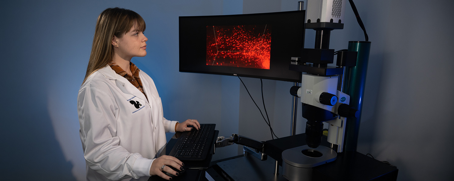
The Brain Health Research Institute (BHRI)’s new light sheet microscope creates 3D imaging of large samples at subcellular resolution (about 200x smaller than the diameter of a human hair). The Blaze, as the device is commonly known, provides an unparalleled ability to image and map complete biological systems from various species, including human samples.
“Kent State currently has the only Blaze light sheet microscope available for use by researchers in Northeast Ohio,” said Aleisha Moore, Ph.D., BHRI assistant director of the Neuroimaging Collaboratory. “Therefore, Kent State researchers have access to an incredibly cutting-edge and versatile microscope that enables them to study physiological systems with unparalleled anatomical detail.”
The microscope offers unmatched imaging speed and, when combined with tissue clearing protocols, rapidly images intact organs. This overcomes the limitations of conventional microscopy, which requires tissue to be sliced into thin sections, and allows users to explore structures at resolutions and scales that traditional methods cannot achieve.

For example, traditional microscopy is like using a single camera to photograph a leaf in a forest, displaying its detailed veins, while a drone-mounted camera captures the entire forest, with the trees appearing blurred. These cameras can’t merge detailed leaf images with the broader landscape. A light sheet microscope, however, allows mapping of the whole forest while zooming in to study individual trees in detail. For neuroscientists, the ability to shift between large-scale and microscopic views is groundbreaking. They can first observe a broad network of neurons and their connections, then focus on the tiny dendritic spines of one cell.
“In my own research, the ability to image entire neural populations and their connections in 3D significantly accelerates our process of identifying the full identity and function of networks that control fertility,” said Moore. “The brain area we study, known as the hypothalamus, contains a diverse mix of cells with complex connections. By imaging these cells in 3D, we can begin to map and understand the cell populations responsible for fertility in ways that traditional microscopy cannot achieve.”
The Blaze was added to the BHRI Collaboratories Lab in 2025 and serves as a valuable resource for research on cell populations in health and disease. One use of the microscope in this facility is rapid imaging of entire brains, which facilitates 3D studies of the healthy brain or disease progression in neurodegenerative conditions such as Alzheimer's disease and cancer. This helps researchers obtain a comprehensive understanding of what the brain should look like, including the changes that occur in a neurodegenerative condition and how new therapies affect disease progression, whether to slow it or cure it. Previously, such detailed data collection took weeks or months, but with Blaze, it now takes only minutes. This speed accelerates research and allows labs to move from studying single “representative” brains to high-throughput experiments with multiple subjects.
As an assistant professor in the Department of Biological Sciences, Moore’s research seeks to identify and understand the neural circuits that are responsible for reproductive capacity. She studies how changes within these networks contribute to reproductive disorders like polycystic ovary syndrome (PCOS). Moore’s grant-funded research also investigates how alterations in brain circuits contribute to reproductive symptoms and related health issues in PCOS, with the goal of finding new treatment options. Understanding the causes of PCOS and developing strategies to prevent and treat it is essential, not only for fertility goals but also because the syndrome poses lifelong health risks including metabolic syndrome, cardiovascular diseases, gynecological cancers, depression, anxiety, eating disorders and more.
"The Blaze microscope is important to my research because it allows for 3D imaging and mapping of the interconnected hypothalamic circuitry that is ultimately responsible for the regulation of mammalian reproduction,” said Alyssa Novak, a fourth year Ph.D. candidate in the Integrative Physiology and Neurobiology program. “This technology will allow me to visualize this circuitry both under physiological conditions and in conditions of infertility, which can provide insights about specific changes in the brain that can alter reproductive function.”
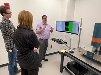
Students and faculty researchers who would like to use the microscope can contact Moore (amoor149@kent.edu) to discuss how to utilize 3D organ imaging in their research, perform optical tissue clearing protocols and receive training.
This innovative technology supports Kent State’s Brain Health Research Institute’s position as a leader in the region, allowing for even greater opportunities to understand the brain, including how it reacts to various diseases and therapies aimed at addressing those conditions.