


Kent State University's College of Public Health and Health Sciences brings together complementary disciplines to support interdisciplinary education and innovation. The U.S. Bureau of Labor Statistics projects a 17% growth in community and social service occupations, including public health roles, from 2022 to 2032.


What Have We Been Up To?

The Elisabeth Severance Prentiss Foundation has awarded $1.75 million to Kent State University’s College of Public Health that will support students and programs, such as the Mobile Flashes program.

The halls of Kent State University are currently echoing with two distinct but harmonized sounds, the rustle of stiff, new white coats and the confident handshakes of departing professionals.
Kent State University’s College of Public Health has earned a national ranking for the university in the newly released U.S.
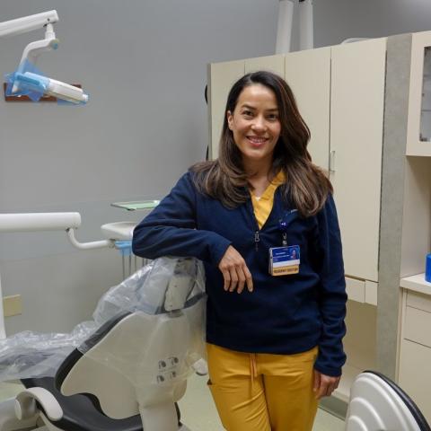
In her first year at MetroHealth's dentistry program, Bonillo Farias is participating in a partnership between MetroHealth and Kent State University that integrates public health education into clinical dental training. The collaboration was made possible by a federal grant awarded to MetroHealth to strengthen dental residents' public health skills.

University Hospitals (UH) has longstanding history of extraordinary support for both Kent State University and the College of Public Health. CPH students work with UH in a variety of ways, including internships, volunteering, and shadowing opportunities.

Catherine Rischar, BSPH, graduated in 2021 with a bachelor’s degree with a minor in psychology from the Kent State University’s College of Public Health.

Limited access to care, lack of cultural and linguistically diverse services, and financial and transportation constraints are only a few of the barriers that hinder many Ohioans from receiving essential dental care.

Image
