


Neuroimaging Collaboratory
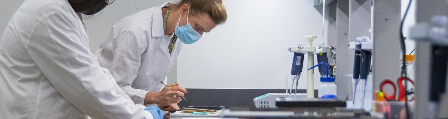
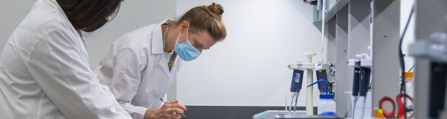
The Neuroimaging Collaboratory provides BHRI members with resources and support for advanced microscopy and image analysis.
To request use of the facilities, please fill out the Application for Use below. Please note that not all fields may apply.
RESOURCES:
- Fluorescence and brightfield microscope with MBF Bioscience
- High throughput Olympus Slide Scanner
- Blaze Light sheet Microscope for 3D imaging of cleared specimens
- Image Analysis Software and Computers: Imaris (Bitplane), Fiji (ImageJ), Deconvolution Software (Olympus), Stereo Investigator (MBF Bioscience), Neurolucida (MBF Bioscience).
Training and Technical Support:
*All users need to complete training on how to use the microscopes and/or computers prior to scheduling the use of equipment.
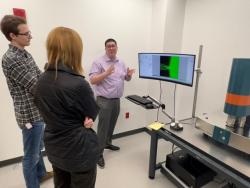
Training and technical support are provided by Dr. Aleisha Moore, BHRI Assistant Director for the Neuroimaging Collaboratory (Blaze Light-sheet microscope, Olympus Slide Scanner and MBF Bioscience Microscope).
Services are also available for the acquisition of pilot data, with imaging and capture conducted by the neuroimaging collaboratory technician.
Scheduling:
After completing training, you will receive access to the Elan Infinity scheduling system.
To schedule use of any of the microscopes or computers, use: https://www.di.kent.edu/facility.
If you have questions about Fusion scheduling or with any questions about navigating the scheduling system, please contact Mary Koch.
User Fees:
We are in the process of developing a user fee-for-service cost structure, along with descriptive material about the Collaboratory, its equipment and facilities, for your use in grant applications.
At the present time, usage is free of charge for all BHRI members.
MICROSCOPES:
Olympus VS200 Research Slide Scanner:
Keywords: high throughput, high speed, fluorescent, brightfield, deconvolution.
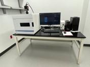
The Slide Scanner is designed to capture high-resolution images of your microscope slides for quantitative analysis. The main advantages of the Slide Scanner are the automation, speed, and ease of capturing large numbers of images. Users can even set up their image-project and leave the Collaboratory to work on other tasks and return later to a full set of images captured automatically.
The slide scanner can capture brightfield and precise fluorescent signals (up to 5 channels: UV, green, red, far red, and near-infrared) with reduced photobleaching. Two slide trays are available for different sizes of microscope slides: 3"x 1" or 3" x 2" slides. A barcode reader automatically captures and records slide information and extensive metadata is available for optimal rigor and reproducibility.
Images can include entire sections or regions of interest within sections. Z-stacks can be captured for the use of deconvolution software (see: Image analysis software). Image analyses can be performed with any software of choice, including Fiji and Imaris (see: Image analysis software). Examples of use: RNA scope or immunofluorescent analysis in multiple experimental groups; full brain analyses of brain area size and neuron morphology.
Technical Details:
VS200 Slide Scanner Olympus
Objectives: 2x, 4x, 20x, and 40x (40x objective requires Immersion Oil)
Slide Trays: 1”x 3” (holds 6 slides) and 2”x 3” (holds 3 slides)
Filter sets for Fluorescence:
- DAPI Filter Set 350/50X, BS400, 460/50M
- EGFP/FITC/CY2 Filter Set 470/40X, BS495, 525/50M
- mCherry/TxRed/Cy3 Filter Set 560/40X, BS585, 630/75M
- CY5 Filter Set 640/30X, BS660, 690/50M
CY7 Filter Set 710/75X, BS760, 810/90M
Fluorescence and bright-field image capturing and analysis:
Keywords: fluorescent, brightfield, whole slide imaging, stereology, morphometric analysis. 

This microscope can be used for a variety of projects, including traditional viewing and capturing images of fluorescent (up to 4 channels: UV, green, red, far red) and brightfield microscope slides. In addition, it contains multiple MBF Bioscience modules that allow for:
- Whole Slide Imaging: Capturing many small high-resolution image tiles or strips and then montaging them to create a full image of a histological section.
- Morphometric and Stereological analyses
MBF Bioscience Module Neurolucida is fully integrated with Leica DMR microscope system hardware to control motorized stage, focus, camera, and optical filters, and is used to capture high quality images and z-stacks of brightfield or fluorescent sections. A key feature is the ability to automatically capture and montage images at high magnification of a large area for high-quality images (whole slide imaging).
Neurolucida is also specifically designed for accurately tracing neuronal structures directly from histological specimens and generating quantitative morphometric data and is used for over 500 morphological quantitative analyses. Analysis can be completed on a separate computer in the collaboratory (see:Image analysis software).
Stereo Investigator provides the means to make accurate, unbiased quantification of the number, length, area, and volume of cells, subcellular and macro structures in tissue specimens. The Stereo Investigator system makes stereology accessible, with easy-to-follow workflows with the most commonly used probes and clear quantitative results. Stereo Investigator can be used for analysis directly on the microscope system, from image stacks, or whole slide images on a separate computer in the Collaboratory (see: Image analysis software).
Examples of use: optimization and validation of antibody staining, single images (or montaged images) of different experimental treatments, dendrite and spine analysis, stereological analysis.
Technical Details:
DMR Leica microscope (Upright)
Objectives: 4x, 10x, 20x, 40x, 60x, 100x (60x and 100x objectives require Immersion Oil)
Slide Holder: 1”x 3” (holds 1 slide) and 2”x 3” (holds 1 slide)
Brightfield, Darkfield, Fluorescence
Monochrome digital camera (Fluorescence) and Color digital camera (Brightfield)
Filter sets for Fluorescence:
- DAPI Filter Set 350/50X, 460/50M
- EGFP/FITC/CY2 Filter Set 470/40X, 525/50M
- mCherry/TxRed/Cy3 Filter Set 560/40X, 630/75M
- CY5 Filter Set 640/30X, 690/50M
UltraMicroscope Blaze light sheet microscope
The Blaze enables rapid 3D visualization of entire biological systems at a subcellular level. This microscope is capable of imaging fluorescent staining in whole brains, organs or tissues from animal models or human samples that have undergone clearing with organic solvent-based methods (e.g., 3DISCO, MACS). Recently, it has been upgraded to enable imaging multiple samples and organs in just minutes.
*Please watch the "Special Webinar: Introduction to Light Sheet Microscopy with the UltraMicroscope Blaze" here: Special Webinar
IMAGE ANALYSIS SOFTWARE AND COMPUTERS:
The Collaboratory also includes two stations equipped with Dell computers loaded with image analysis software. These software packages can be used with images captured on the microscopes in the Collaboratory or other microscopes including any confocal or bright-field/fluorescent microscope:
- Imaris (Bitplane) Imaris is the market-leading software for visualization and analysis of images collected from wide-field, confocal (including spinning disk), light sheet, two-photon, electron microscopy, CLEM, OPT, and other microscopes, with over 4,000 publications in 2020 alone.
- Fiji (ImageJ)
- Deconvolution software (Olympus)
- Stereo Investigator (MBF Bioscience)
- Neurolucida (MBF Bioscience)
CONTACT US:
To schedule a consultation on study design, use of instrumentation, or image analysis or for technical support, please email:
Dr. Aleisha Moore: amoor149@kent.edu