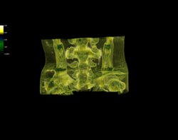
Figure 1

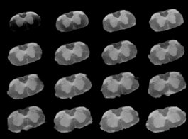
Figure 2
The 500 MHz NMR spectrometer in the Department of Chemistry and Biochemistry has recently been upgraded with new, digital control console and new software. The new electronics offer greater stability for long experiments and new software has an extensive library of pulse sequences and data processing tools. An additional, non-standard feature of this instrument is its micro-imaging capability (micro-MRI or NMR microscopy), which can be used for generating high-quality MR images with 10 microns spatial resolution.
The acquisition and installation of the new console was made possible by a grant of $327,753 from the National Science Foundation (CHE-1048645). Professor Tubergen is the project Principal Investigator (PI), and co-PI's include Professors Brasch, Huang, Khitrin, and Kooijman (Biological Sciences). A new gradient probe with ultra-strong magnetic field gradient is also being acquired to support NMR measurements of diffusion rates.
Professor Khitrin, in collaboration with Rob Clements (Biological Sciences), uses micro-imaging capabilities of this NMR spectrometer to develop new MRI techniques. The examples below show 3D reconstruction of a fragment of a mouse embryo (Figure 1) and slices of a mouse spinal cord (Figure 2). The diameter of both objects is about 2mm.
